Need urgent care? 24/7 Mobile & After-Hours Emergency Physiotherapy Call (02) 8111 5633 For life-threatening emergencies, always call 000
Knee

Patellar Dislocation

5.0 average rating on Google
Registered NDIS Provider
SIRA Registered Provider

You planted your foot, turned, and your kneecap shot out to the side of your knee. Maybe it slid back in as soon as you straightened your leg. Maybe someone in the emergency department put it back for you. Either way, the knee has swollen up, it's tender along the inner edge of the kneecap, and the question on your mind is whether it's going to happen again. If this is your teenager's knee, you're in very familiar territory: most of these injuries happen to active 10- to 17-year-olds 1.

What's actually causing it

Your kneecap (the patella) sits in a shallow groove at the lower end of your thigh bone and glides up and down in it as your knee bends. On its inner side sits the medial patellofemoral ligament, or MPFL, one of the soft tissues that keep it in that groove.

The typical dislocation happens when your foot is firmly planted, your knee is pushed inwards and your thigh bone rotates inwards over the top of it. The kneecap is left behind and slips out to the side 1. In real life that looks like a netball landing, a pivot at footy training, or a slip that twists the knee. When the kneecap goes, the MPFL is injured 3. The kneecap can also scrape the joint's cartilage, or break off a small piece of bone and cartilage (an osteochondral fracture) from either the kneecap or the thigh bone 1.

Why do some knees dislocate and others don't? A few things make it more likely: an MPFL that's already been damaged, a kneecap that's unusually shaped or positioned, a shallow groove, or loose, hypermobile joints 1. Growth plates that are still open and generally flexible ligaments add to the risk. So do some things physiotherapy can work on, such as an underdeveloped inner quadriceps muscle (the VMO) or outer thigh muscles that overpower it 2.

Whether girls and women are more affected is disputed. One Australian guideline finds no clear difference between the sexes 1. Another guideline and a Cochrane review suggest it's more common in females 2,3, possibly because of limb alignment and looser joints 4.
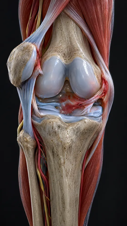

Front of the knee showing the kneecap slipped to the outer side and the torn ligament on the inner side

Dislocated kneecap or ACL? How to tell the difference

Plenty of people arrive with a swollen, painful knee and nothing visibly out of place, because the kneecap popped back in by itself 1. A twist, a sense that something shifted, then fast swelling is also the classic story of an ACL injury. The overlap is real: a patellar dislocation is the second most common cause of bleeding into the knee joint, behind only ACL injury 1.

Some things point towards the kneecap. You may have seen it sitting out to the side, with the front of the knee looking flat and a bulge on the outer edge. The knee usually stays bent while the kneecap is still out. Afterwards, there's tenderness along the inner border of the kneecap 1.

Neither injury rules out the other, and the other ligaments need checking once the kneecap is back in place 1. That's a job for a hands-on examination, not guesswork.

How it's diagnosed

Often the kneecap is already back in place by the time anyone examines the knee, so the diagnosis comes from your story, a hands-on examination and imaging together 1 2. The story that fits is a twist, a knee that looked out of shape, then one that corrected itself 1. On examination the clinician may gently glide the kneecap outwards. This is the apprehension test. A kneecap that moves a long way suggests the soft tissues on its inner side aren't holding it properly 2.

X-rays are taken from three angles once the kneecap is back in. They aren't really there to check its position. They're looking for small chips of bone, because even a small one can mean a larger piece of cartilage has come away 1.

How we treat it

The first days usually happen elsewhere: emergency or your GP, X-rays, a brace and an MRI. Our part begins after that. For a first dislocation with no bone or cartilage damage, the guideline pathway is physiotherapy once the brace comes off. The program focuses on getting movement back, building strength (particularly the VMO), and retraining balance and joint position sense, known as proprioception 1.

1
While you're still in the brace. Straight-leg raises in the splint help stop the quadriceps wasting away 1.
2
A staged program through sports physiotherapy. A NSW children's hospital guideline sets out four phases: initial, basic function, advanced function and return to sport. You move up by passing criteria, not by how many weeks have gone by 2. For example, before the final phase you need more than 25 single-leg sit-to-stands with good knee alignment, plus a balance-reach score at least 95% of your other leg's 2.
3
Return-to-sport testing. Hop tests have to reach more than 95% of your uninjured side before you're cleared 2. We measure this rather than guess, because six months after injury the injured leg is still commonly only about 80% as strong as the other one 2.
4
Patience before surgery. Where no fragment needs fixing, the guideline recommends at least six months of physiotherapy. Surgery is considered if the kneecap dislocates three or more times during or after that time 2.
5
Rehabilitation after an operation. If your MRI shows a fragment that needs fixing 1, post-surgical rehabilitation follows the same logic, staged around your surgeon's protocol.

Other treatments we may use

Bracing, and how long for.

The two Australian guidelines disagree on this. One recommends about three weeks in a brace locked straight 1. The more recent physiotherapy guideline says no longer than two weeks, and that "there is no evidence to support bracing beyond this period" 2. The doctor managing your injury sets this, and we work to their plan.

Taping or a kneecap stabilising brace.

Longer term, a brace that blocks the kneecap from moving outwards can replace taping 2. The guideline lists it as an option but doesn't measure how much it reduces re-dislocation. So we use it for support as you return to sport, never as a substitute for strength.

Manual therapy.

Hands-on techniques can help ease stiffness once the brace comes off. None of the evidence we've drawn on tests manual therapy specifically for patellar dislocation. That makes it an extra at most, not the main treatment.

Surgery.

An operation is needed when the MRI shows a significant loose piece of cartilage or bone 1. Reconstructing the torn soft tissues straight after a first dislocation usually isn't needed, as it doesn't appear to change the long-term outcome 1. For surgery versus non-surgical care more broadly, the evidence is unsettled. A 2023 review of 10 trials found that surgery might mean fewer repeat dislocations. But all of the evidence was very low certainty, it was unclear whether surgery improved knee function, and satisfaction was similar either way 3. That decision belongs to you and an orthopaedic surgeon.

Your recovery path: Reset, Rebuild, Return

After a kneecap dislocation, rehabilitation follows The Well Motion Recovery Path™. Your knee earns each step up by passing tests, whatever the date.

  • Reset: the brace period, set by your doctor 1,2. Straight-leg raises in the splint 1, with physiotherapy starting inside the first month 7. Expect a swollen knee and a thinner thigh on that side. We move you on once the brace is off and you can walk without the pain building.
  • Rebuild: the longest phase. Movement, VMO strength and balance work 1, with manual therapy if bracing has left the knee stiff. Expect the knee to feel untrustworthy on turns after the pain has gone. We move you on when you pass the sit-to-stand and balance-reach criteria 2 or, if sport is not your aim, when the knee does what your daily tasks ask of it.
  • Return: sport, work, or stairs taken without thinking. We run hop tests against your other leg 2. You leave with a long-term exercise program 2, plus taping or a stabilising brace if you need support 2.

How long it usually takes. A panel of specialists who treat adolescents put return to sport at 2 to 4 months after a first dislocation. Only 68% of them agreed, and it is expert opinion, not measured outcomes 7. At six months the injured leg is commonly still about 80% as strong as the other 2. If you need surgery, your surgeon's protocol sets the timeline.

Reducing the risk of it happening again

The best exercise program after a dislocation is still unknown 6. What has been measured is who dislocates again:

  • Know your own odds. Pooling 17 studies, about one in three people dislocated again. Younger age, open growth plates, a shallow groove and a high-riding kneecap each raised the risk. With none of the risk factors, the repeat rate was 7.7 to 13.8%. With three, it was 70.4 to 78.5% 5.
  • Work on what exercise can change. An underdeveloped inner quadriceps and outer thigh muscles that overpower it are the factors the physiotherapy guideline calls amenable to treatment. The shape of the groove and the height of the kneecap aren't 2.
  • Finish the rehab, then keep it up. The guideline's aim is equal strength in both legs, and it raises consistent, long-term exercise as something to discuss 2.
  • Be realistic about anatomy. People with no or mild risk factors do predictably well with rehabilitation. Those with major alignment problems tend to do poorly even when they stick to it 2. If that's your knee, ask about an orthopaedic opinion.

When to get it checked properly

  • The kneecap is still out of place. Call us. It should be put back by a clinician, by gently straightening the knee 1. We assess it and manage it, and get you to hospital when that's what it needs.
  • It's your first dislocation, even if it went straight back in. Book an assessment with us within three days. An MRI within about seven days is recommended 1, and we'll tell you how to arrange it.
  • The knee swelled up large and tight within hours. That makes a bone or cartilage injury more likely 1.
  • The knee locks or catches. A loose fragment inside the joint can cause this 1.
  • Pain isn't settling with regular pain relief and the splint 1.
  • The other kneecap has dislocated before. That raises the risk six-fold and warrants an orthopaedic referral 1.
  • It keeps happening despite several months of good physiotherapy 1.

None of this means the worst has happened. Most of the time it means getting the right information early, so your rehabilitation is built on it.

Your first appointment

We start by listening: how it began, what makes it worse, and what it's stopping you from doing. Then we examine the area, explain what we find in plain English, and agree a plan with you. Your first visit is 40 minutes at Engadine and 30 minutes at our other clinics.

We see patients at Engadine, Mount Annan, Narellan and Appin, and at home through our mobile physiotherapy service. If you have private health cover, here's how health fund rebates work for physiotherapy.

FAQs

How do you fix a patellar dislocation?

First the kneecap is put back, usually by gently straightening the knee with pressure on the side of the kneecap 1. Then comes a short period in a brace, an MRI to check for bone or cartilage damage, and a physiotherapy program for movement, strength and balance 1,2. Surgery is kept for loose fragments or repeated dislocations 1,2.

How serious is a patella dislocation?

More serious than it often feels once the kneecap is back in. Bone or cartilage fractures occur in 25 to 75% of cases 1. After the first dislocation, a knee is seven times more likely to dislocate again 2.

Can you fully recover from a dislocated patella?

Many people do, but an honest answer includes the risks. Over a third of people end up with recurrent instability. The children's hospital guideline also reports that nearly half never return to sport because the knee keeps feeling unstable 1. Ongoing pain and early wear behind the kneecap are reported too 2. That's the case for criteria-based rehabilitation: progressing on measured strength, balance and hop tests rather than the calendar 2.

My kneecap popped out of place. What should I do?

Don't force it back yourself. Call us if it's still out. If it has gone back in, book an assessment with us within three days; an MRI within about seven days is recommended 1.

Is a dislocated kneecap worse than a torn ACL?

They're different injuries rather than better or worse versions of one another. A dislocated kneecap injures the MPFL 3 and can damage the cartilage 1. An ACL injury involves a different ligament. Both commonly cause bleeding into the knee 1, and a proper examination is how you find out which one you're dealing with.

More Knee conditions

Knee

Meniscus Tear

Catching, locking or swelling after a twist, sometimes with a knee that won't fully straighten.

Read more
Knee

MCL Injury

The MCL runs down the inside of your knee. Most injuries happen when something hits the outside of the knee and forces it inwards; it's the ligament most commonly injured in knee trauma.

Read more
Knee

Prepatellar Bursitis

A bursa is a small fluid-filled sac that cushions bone from the soft tissue around it. The prepatellar bursa sits at the front of your knee, over the kneecap. When it's irritated it swells, and a tender, warm swelling right over the bursa is the classic sign.

Read more
Knee

Baker's Cyst

A Baker's cyst is a build-up of fluid behind the knee, usually excess fluid from the knee joint itself.

Read more

If your kneecap has come out and nobody has yet explained what the next six months involve, or you're already rehabbing and don't know whether you're on track, book an appointment at Engadine, Mount Annan, Narellan or Appin.

We'll build the program around where your knee actually is, with real tests to pass rather than dates to wait out. If you're not sure the kneecap is the problem, start from the Knee section of our Injury Finder instead.

Book an Assessment
Ahmed Elsayed, Principal Physiotherapist
Reviewed by Ahmed Elsayed Principal Physiotherapist at Well Motion

References

  1. Patellar Dislocation: Emergency Department. Clinical Practice Guidelines, The Royal Children's Hospital Melbourne. Last updated October 2020. https://www.rch.org.au/clinicalguide/guideline_index/fractures/Patellar_Dislocation_-_Emergency_Department/
  2. Patella Dislocation: Physiotherapy Management: CHW. Guideline 2022-137, Sydney Children's Hospitals Network (NSW Health), 2022. https://resources.schn.health.nsw.gov.au/policies/policies/pdf/2022-137.pdf
  3. Smith TO, et al. Surgical versus non-surgical interventions for treating patellar dislocation. Cochrane Database of Systematic Reviews, 2023. PMID 36692346. https://doi.org/10.1002/14651858.CD008106.pub4
  4. Parikh SN, Lykissas MG, Gkiatas I. Predicting risk of recurrent patellar dislocation. Current Reviews in Musculoskeletal Medicine, 2018;11(2):253-260. PMID 29736871. https://pmc.ncbi.nlm.nih.gov/articles/PMC5970115/
  5. Huntington LS, Webster KE, Devitt BM, Scanlon JP, Feller JA. Factors associated with an increased risk of recurrence after a first-time patellar dislocation: a systematic review and meta-analysis. American Journal of Sports Medicine, 2020;48(10):2552-2562. PMID 31825650. https://doi.org/10.1177/0363546519888467
  6. Forde C, Haddad M, Hirani SP, Keene DJ. Is an individually tailored programme of intense leg resistance and dynamic exercise acceptable to adults with an acute lateral patellar dislocation? A feasibility study. Pilot and Feasibility Studies, 2021;7(1):197. PMID 34749823. https://doi.org/10.1186/s40814-021-00932-x
  7. Parikh SN, Schlechter JA, Veerkamp MW, Stacey JD, Gupta R, Pendleton AM, Shea KG, Friel NA, Molony JT, Yaniv M, Rhodes J, Finlayson CJ, Williams BA, Ellington M. Consensus-based guidelines for management of first-time patellar dislocation in adolescents. Journal of Pediatric Orthopedics, 2024;44(4):e369-e374. PMID 38258884. https://doi.org/10.1097/BPO.0000000000002616